Bioassay report: "La LDL "mínimamente modificada" (LDLmm) induce en las células endoteliales la expresión de moléculas de adhesión, proteínas quimiotácticas y factores de crecimiento que dan lugar al reclutamiento de monocitos y linfocitos hacia la pared vascular a través de interacciones específicas entre las molécula ...s de adhesión endoteliales y las integrinas de aquéllos.".... Its True!
JC teacher
miércoles, 30 de diciembre de 2009
jueves, 24 de diciembre de 2009
Como dice el Dr Cann: The origins of cavity-causing bacteria

Bifidobacteria are relatively abundant inhabitants of the gastrointestinal tract of humans and animals. Many bifidobacterial species, in conjunction with other members of the intestinal microbiota are believed to contribute to host nutrition, while also impacting on intestinal pH, cell proliferation and differentiation, development and activity of the immune system, and innate and acquired responses to pathogens. These perceived beneficial health effects have driven commercial exploitation of bifidobacteria as live components of many functional foods and therapeutic adjuncts. However, bifidobacteria have also been isolated from the human oral cavity, where their presence is linked to the progression of tooth decay: bifidobacteria have been detected in high numbers in infected dentine from carious lesions in children and have been associated with childhood dental caries. can be found as part of the microbiota implicated in human dental caries. In recent surveys of oral bifidobacteria associated with caries in adults and children and root caries in adults, Bifidobacterium dentium was the most frequently isolated Bifidobacterium species, representing approximately eight percent of the culturable bacteria isolated from active lesions. This species is capable of acidogenesis to produce a final pH in glucose-containing media below pH 4.2, sufficient to cause extensive demineralisation of tooth tissues. B. dentium may therefore significantly contribute to the pathogenesis of dental caries which is one of the most common chronic diseases, remaining untreated in many underdeveloped countries where dental pain is often alleviated only by the loss or extraction of the affected tooth.
Researchers have now uncovered the complete genetic make-up of the cavity-causing bacterium B. dentium Bd1, revealing the genetic adaptations that allow this microorganism to live and cause decay in the human oral cavity. Bifidobacteria, largely known as long-term beneficial gut bacteria, are often included as probiotic components of food to aid digestion and boost the immune system. However, not all species within the genus Bifidobacterium provide beneficial effects to the host’s health. In fact, B. dentium is an opportunistic pathogen since it has been linked to the development of tooth decay. The genome sequence of B. dentium Bd1 reveals how this microorganism has adapted to the oral environment through specialized nutrient acquisition features, acid tolerance, defences against antimicrobial substances and other gene products that increase fitness and competitiveness within the oral niche. This report identifies, through various genomic approaches, specific adaptations of a Bifidobacterium taxon to a lifestyle as a tooth decay-causing bacterium. The data in this study indicate that the genome of this opportunistic pathogen has evolved through only a small number of horizontal gene acquisition events, highlighting the narrow boundary that separates bacteria that are long-term residents on or in the human body from opportunistic pathogens.
The Bifidobacterium dentium Bd1 Genome Sequence Reflects Its Genetic Adaptation to the Human Oral Cavity. 2009 PLoS Genet 5(12): e1000785. doi:10.1371/journal.pgen.1000785
Bifidobacteria, one of the relatively dominant components of the human intestinal microbiota, are considered one of the key groups of beneficial intestinal bacteria (probiotic bacteria). However, in addition to health-promoting taxa, the genus Bifidobacterium also includes Bifidobacterium dentium, an opportunistic cariogenic pathogen. The genetic basis for the ability of B. dentium to survive in the oral cavity and contribute to caries development is not understood. The genome of B. dentium Bd1, a strain isolated from dental caries, was sequenced to completion to uncover a single circular 2,636,368 base pair chromosome with 2,143 predicted open reading frames. Annotation of the genome sequence revealed multiple ways in which B. dentium has adapted to the oral environment through specialized nutrient acquisition, defences against antimicrobials, and gene products that increase fitness and competitiveness within the oral niche. B. dentium Bd1 was shown to metabolize a wide variety of carbohydrates, consistent with genome-based predictions, while colonization and persistence factors implicated in tissue adhesion, acid tolerance, and the metabolism of human saliva-derived compounds were also identified. Global transcriptome analysis demonstrated that many of the genes encoding these predicted traits are highly expressed under relevant physiological conditions. This is the first report to identify, through various genomic approaches, specific genetic adaptations of a Bifidobacterium taxon, Bifidobacterium dentium Bd1, to a lifestyle as a cariogenic microorganism in the oral cavity. In silico analysis and comparative genomic hybridization experiments clearly reveal a high level of genome conservation among various B. dentium strains. The data indicate that the genome of this opportunistic cariogen has evolved through a very limited number of horizontal gene acquisition events.
Tomado de :
Cann Alan, MicrobiologyBytes, (on line) url: http://www.microbiologybytes.com/blog/2009/12/24/the-origins-of-cavity-causing-bacteria/?utm_source=feedburner&utm_medium=feed&utm_campaign=Feed%3A+Microbiologybytes+%28MicrobiologyBytes%29 (Cited on Dec 24/09)
miércoles, 9 de diciembre de 2009
Microbiologia entomologica y vida en sociedad....
Les comparto un interesante dato publicado por Manolo en su blog "curiosidades de la microbiologia":
JC
JC
Los superpoderes de las cucarachas  Hay una vieja historia que dice que en caso de guerra nuclear total, los únicos supervivientes serían las cucarachas. Lo cierto es que uno no puede dudarlo si lee algo sobre lo que son capaces de hacer. El último de sus "superpoderes" ha sido descrito en un reciente artículo publicado en la revista PNAS a cargo de un grupo investigador liderado por la doctora Nancy Moran.
En la escuela aprendemos que el nitrógeno es un gas inerte y que es el principal componente de la atmósfera. Lo de "inerte" tiene su importancia, porque si fuera reactivo como el oxígeno la vida en el planeta sería muy distinta de como la conocemos ahora. El nitrógeno gaseoso (N2) no puede ser usado por casi ninguna célula. Es una molécula tremendamente estable, y es necesario romperla para poder combinar el nitrógeno con otros elementos para así sintetizar proteínas y ácidos nucleicos entre otras biomoléculas.
Son muy pocos los seres vivos que consiguen romper esa molécula de N2, y todos ellos son microorganismos. Son los llamados fijadores del nitrógeno. Quizás el más famosos de ellos sea la bacteria Rhizobium melliloti, que consigue establecer una simbiosis con plantas leguminosas para poder realizar el proceso de fijación. Su simbiosis podría resumirse de la siguiente forma: la planta da cobijo y alimento a la bacteria y ésta a su vez consigue fijar nitrógeno gaseoso en sus compuestos orgánicos, que luego cede a la planta. Una vez ya tenemos el nitrógeno en un compuesto orgánico ya podemos imaginarnos que llega a los animales a través de la dieta: la planta es comida por un herbívoro, y éste a su vez es comido por un carnívoro, etc.
Pero a veces hay problemas con el suministro de nitrógeno orgánico. Así que muchos seres vivos intentan establecer simbiosis con microorganismos que metabolicen eficientemente el nitrógeno y tener un problema alimentario menos del que preocuparse. Y ese truco parece que también lo han aprendido las cucarachas, unos animalitos acostumbrados a alimentarse de cualquier cosa.
Como muchos otros insectos, las cucarachas excretan nitrógeno en forma de ácido úrico, pero con una sutil diferencia. Lo excretan al interior de su cuerpo, en concreto a los llamados cuerpos grasos. De esa forma pueden recuperarlo en caso de necesidad. Pero ¿cómo pueden reciclar ese ácido úrico? Hay dos tipos de células en dichos cuerpos grasos. Uno son adipocitos en los que se acumulan grasas y cristales de ácido úrico. El otro tipo celular consiste en los micetocitos o bacteriocitos, que están llenos de bacterias. En concreto se trata de una bacteria Gram negativa de nombre Blattabacterium y que se transmite de manera vertical entre estos insectos. Células de Blattabacterium en el interior de un micetocito. Las bacterias están señaladas con las flechas blancas. El núcleo del micetocito tiene color azul y están señalados por la flecha blanca punteada (Fuente PNAS).
El grupo investigador ha secuenciado el genoma de la bacteria que se encuentra dentro de la cucaracha Periplaneta americana. Han confirmado que pertenece a las Flavobacterias y que su "pariente más próximo" es el endosimbionte Sulcia muelleri, que vive dentro de insectos especializados en chupar la savia. Mediante la información genómica contenida en sus 636 Mb han reconstruido el posible metabolismo de Blattabacterium y han encontrado que puede reciclar el nitrógeno a partir de urea o de amonio ya que posee genes para la ureasa y la glutamato deshidrogenasa, aunque carece de enzimas uricolíticas. Además es capaz de producir aminoácidos esenciales, varias vitaminas y otros compuestos útiles para la cucaracha. También han concluido que la asociación entre Blattabacterium y las cucarachas se originó hace unos 140 millones de años, en pleno Jurásico.
 Predicción del metabolismo de Blattabacterium. En azul se indican las rutas para aminoácidos, en verde la de metabolitos varios y en púrpura la de las vitaminas. En rojo se indican los aminóacidos no producidos por la bacteria y que son originados por la cucaracha(Fuente PNAS).
Como no se han encontrado genes para enzimas uricolíticas en el genoma de Blattabacterium no está muy claro el modo en que aprovechan el ácido úrico. Los investigadores proponen tres hipótesis. La primera es que la actividad uricolítica está producida por otra enzima no identificada en Blattabacterim. La segunda es que la actividad uricolítica sea debida a las células de la cucaracha. Pero en contra de esta hipótesis está el hecho de que si se trata a las cucarachas con antibióticos la actividad uricolítica desaparece. La tercera es que la cucaracha acumule el úrico en los adipocitos y cuando necesita nitrógeno, transporta el úrico al intestino donde la microflora intestinal es capaz de descomponerlo en amonio, que vuelve a ser absorbido y transportado por la hemolinfa a los micetocitos.
Sea lo que sea, hay que reconocer que las cucarachas tienen un kit de supervivencia que sería la envidia de McGyver.
Hay una vieja historia que dice que en caso de guerra nuclear total, los únicos supervivientes serían las cucarachas. Lo cierto es que uno no puede dudarlo si lee algo sobre lo que son capaces de hacer. El último de sus "superpoderes" ha sido descrito en un reciente artículo publicado en la revista PNAS a cargo de un grupo investigador liderado por la doctora Nancy Moran.En la escuela aprendemos que el nitrógeno es un gas inerte y que es el principal componente de la atmósfera. Lo de "inerte" tiene su importancia, porque si fuera reactivo como el oxígeno la vida en el planeta sería muy distinta de como la conocemos ahora. El nitrógeno gaseoso (N2) no puede ser usado por casi ninguna célula. Es una molécula tremendamente estable, y es necesario romperla para poder combinar el nitrógeno con otros elementos para así sintetizar proteínas y ácidos nucleicos entre otras biomoléculas.
Son muy pocos los seres vivos que consiguen romper esa molécula de N2, y todos ellos son microorganismos. Son los llamados fijadores del nitrógeno. Quizás el más famosos de ellos sea la bacteria Rhizobium melliloti, que consigue establecer una simbiosis con plantas leguminosas para poder realizar el proceso de fijación. Su simbiosis podría resumirse de la siguiente forma: la planta da cobijo y alimento a la bacteria y ésta a su vez consigue fijar nitrógeno gaseoso en sus compuestos orgánicos, que luego cede a la planta. Una vez ya tenemos el nitrógeno en un compuesto orgánico ya podemos imaginarnos que llega a los animales a través de la dieta: la planta es comida por un herbívoro, y éste a su vez es comido por un carnívoro, etc.
Pero a veces hay problemas con el suministro de nitrógeno orgánico. Así que muchos seres vivos intentan establecer simbiosis con microorganismos que metabolicen eficientemente el nitrógeno y tener un problema alimentario menos del que preocuparse. Y ese truco parece que también lo han aprendido las cucarachas, unos animalitos acostumbrados a alimentarse de cualquier cosa.
Como muchos otros insectos, las cucarachas excretan nitrógeno en forma de ácido úrico, pero con una sutil diferencia. Lo excretan al interior de su cuerpo, en concreto a los llamados cuerpos grasos. De esa forma pueden recuperarlo en caso de necesidad. Pero ¿cómo pueden reciclar ese ácido úrico? Hay dos tipos de células en dichos cuerpos grasos. Uno son adipocitos en los que se acumulan grasas y cristales de ácido úrico. El otro tipo celular consiste en los micetocitos o bacteriocitos, que están llenos de bacterias. En concreto se trata de una bacteria Gram negativa de nombre Blattabacterium y que se transmite de manera vertical entre estos insectos.
Células de Blattabacterium en el interior de un micetocito. Las bacterias están señaladas con las flechas blancas. El núcleo del micetocito tiene color azul y están señalados por la flecha blanca punteada (Fuente PNAS).El grupo investigador ha secuenciado el genoma de la bacteria que se encuentra dentro de la cucaracha Periplaneta americana. Han confirmado que pertenece a las Flavobacterias y que su "pariente más próximo" es el endosimbionte Sulcia muelleri, que vive dentro de insectos especializados en chupar la savia. Mediante la información genómica contenida en sus 636 Mb han reconstruido el posible metabolismo de Blattabacterium y han encontrado que puede reciclar el nitrógeno a partir de urea o de amonio ya que posee genes para la ureasa y la glutamato deshidrogenasa, aunque carece de enzimas uricolíticas. Además es capaz de producir aminoácidos esenciales, varias vitaminas y otros compuestos útiles para la cucaracha. También han concluido que la asociación entre Blattabacterium y las cucarachas se originó hace unos 140 millones de años, en pleno Jurásico.
Predicción del metabolismo de Blattabacterium. En azul se indican las rutas para aminoácidos, en verde la de metabolitos varios y en púrpura la de las vitaminas. En rojo se indican los aminóacidos no producidos por la bacteria y que son originados por la cucaracha(Fuente PNAS).Como no se han encontrado genes para enzimas uricolíticas en el genoma de Blattabacterium no está muy claro el modo en que aprovechan el ácido úrico. Los investigadores proponen tres hipótesis. La primera es que la actividad uricolítica está producida por otra enzima no identificada en Blattabacterim. La segunda es que la actividad uricolítica sea debida a las células de la cucaracha. Pero en contra de esta hipótesis está el hecho de que si se trata a las cucarachas con antibióticos la actividad uricolítica desaparece. La tercera es que la cucaracha acumule el úrico en los adipocitos y cuando necesita nitrógeno, transporta el úrico al intestino donde la microflora intestinal es capaz de descomponerlo en amonio, que vuelve a ser absorbido y transportado por la hemolinfa a los micetocitos.
Sea lo que sea, hay que reconocer que las cucarachas tienen un kit de supervivencia que sería la envidia de McGyver.
Microbiologia en Accion - Curiosidades...
 LIPIDO ASESINO....
 (Para continuar la lectura seguir este vìnculo):
http://curiosidadesdelamicrobiologia.blogspot.com/2009/12/lipido-asesino.html
JC
domingo, 6 de diciembre de 2009
Juan C Gonzalez has shared a presentation on Slideshare
Check out this cool presentation on SlideShare:
Title: "Social Media Monitoring & Measurement 09.22.09"
Link: http://www.slideshare.net/kellykearney/ku-masters-presentation-jour-831-technology-in-marketing-communications-092209
Juan C Gonzalez
--
Not on SlideShare yet? Get a free account here.
SlideShare is the world's largest community for sharing presentations on the web. And it is REALLY USEFUL - you can view, share and download presentations on virtually any topic under the sun.
jueves, 3 de diciembre de 2009
Sus exámenes Finales...
Ya están calificados, estaré en el Centro de Computo junto a Rectoría en la mañana, como entre las 10 y 11 a.m. Y luego, en el Centro de IDiomas, Salón B1 - de 1:30 a 5:00 p.m.
Por favor recogerlos para que puedan retroalimentar su proceso....
JC Teacher
Por favor recogerlos para que puedan retroalimentar su proceso....
JC Teacher
lunes, 30 de noviembre de 2009


sábado, 21 de noviembre de 2009
Evaluación Final del Curso

Recuerden revisar los temas que discutimos en clase para su evaluación final....
Pronto les aviso el salón que nos han asignado
JC


viernes, 20 de noviembre de 2009
Control Biológico

Universidad Católica de Oriente - Facultad de Educación
Curso: Microbiología – Sesión # 17
Docente: Esp. Juan Carlos González Sánchez
Noviembre 21 de 2009
Tópico: Recopilación Sesión Protozoarios - Control Biológico
1. Recuento sesión anterior: Protozoarios.
2. Control Biológico.
2.1. Concepto
2.2. Estrategias
2.3. Historia
Leemos el siguiente texto, se elabora texto resumen:
http://es.wikipedia.org/wiki/Control_biol%C3%B3gico
Revisión del siguiente resumen:
http://www.pv.fagro.edu.uy/fitopato/cursos/fitopato/practicas/cbiologico.html
Teacher JC
Facultad de Educación Universidad Católica de Oriente
viernes, 13 de noviembre de 2009


Sesión Noviembre 14/09
Universidad Católica de Oriente - Facultad de Educación
Curso: Microbiología – Sesión # 16
Docente: Esp. Juan Carlos González Sánchez M.D.
Noviembre 14 de 2009
Tópico: Protozoos
1. Generalidades de los protozoarios
2. Ciclo de Vida de un protozoario
3. Ejemplo de protozoarios de importancia clínica
1. Generalidades de los Protozoos:
Leemos el siguiente texto y elaborar texto resumen:
http://www.alaquairum.net/generalidades_protozoos.htm
2. Ciclo de vida de un protozoo:
http://vanguardia.udea.edu.co/cursos/cursos%202005-2/Parasitologia/maestre.ppt
Diapositivas 1, 2 y 3.
3. Ejemplos de protozoarios de importancia clínica.
Consultar el ciclo de vida y la etiología (cómo se comporta la patología) del Plasmodium, Toxoplasma y Tripanosoma.
Ejemplo: www.santacruztiene.com/archivos/toxoplasmosis.ppt
4. Trabajo Autónomo:
4.1 Se discutirá los hallazgos en la próxima sesión.
4.2 Elaborar una entrada en su blog o portafolio donde se de cuenta de las generalidades (actividad 1) y se presente ciclo de vida de uno de los protozoos (actividad 3)ç
Teacher JC
Facultad de Educación
Universidad Católica de Oriente

viernes, 6 de noviembre de 2009
ALGAS
Para la revisión del tema de Algas, chequear estos dos links, y con base en ellos elaborar un documento resumen, el cual debe incluir algunas imágenes que permitan una mejor comprensión del tema.
1. http://www2.uah.es/botanica_bio/descargas/miriamteoria07-08/ALGAS/procariotas.pdf
2. http://www.librosvivos.net/smtc/files/las_algas.pdf
Teacher JC
1. http://www2.uah.es/botanica_bio/descargas/miriamteoria07-08/ALGAS/procariotas.pdf
2. http://www.librosvivos.net/smtc/files/las_algas.pdf
Teacher JC
martes, 27 de octubre de 2009
Metabolism, cell growth and the bacterial cell cycle

The life of a bacterial cell is feast or famine. To survive the bacterium must rapidly adapt to changing environmental conditions. Colonization of the mammalian gut provides an enteric organism with an abundant source of carbohydrates, whereas a flash flood instantly depletes the nutrient supply for a soil bacterium. Nutrient-rich conditions lead to a decrease in mass doubling time and an increase in cell size, whereas nutrient-poor conditions curtail growth and reduce cell size. Changes in growth rate must be accompanied by changes in the cell cycle to ensure that cell division stays coordinated with mass doubling, chromosome replication and chromosome segregation. How organisms adjust their cell cycle dynamics to compensate for changes in nutritional conditions is an important outstanding question in bacterial physiology. Recent work suggests that multiple signalling pathways transmit nutritional and growth rate information directly to the cell cycle machinery. Multiple signalling pathways permit cells to constantly sample their environments and fine-tune cell cycle processes, a substantial advantage under challenging conditions.
Adaptation to fluctuations in nutrient availability is a fact of life for single-celled organisms in the ‘wild’. A decade ago our understanding of how bacteria adjust cell cycle parameters to accommodate changes in nutrient availability stemmed almost entirely from elegant physiological studies completed in the 1960s. This article summarizes recent groundbreaking work in this area and discuss potential mechanisms by which nutrient availability and metabolic status are coordinated with cell growth, chromosome replication and cell division.
Metabolism, cell growth and the bacterial cell cycle. Nature Reviews Microbiology 7, 822 (2009)
Related:
Bacterial Chemoreceptors
A day in the life of a cyanobacterium
How smart are bacteria?
Tomado de: http://www.microbiologybytes.com/blog/2009/10/20/metabolism-cell-growth-and-the-bacterial-cell-cycle/ citado en 27 Oct 2009.
domingo, 25 de octubre de 2009

endotelinas sent you a video: "Protozoarios 2"
| | help center | e-mail options | report spam |
| endotelinas has shared a video with you on YouTube: Protozoarios - 2 parte | |
| © 2009 YouTube, LLC 901 Cherry Ave, San Bruno, CA 94066 | |
endotelinas sent you a video: "Protozoo"
| | help center | e-mail options | report spam |
| endotelinas has shared a video with you on YouTube: Protozoos - Introduction | |
| © 2009 YouTube, LLC 901 Cherry Ave, San Bruno, CA 94066 | |
endotelinas sent you a video: "Protozoarios 1"
| | help center | e-mail options | report spam |
| endotelinas has shared a video with you on YouTube: Los protozoarios | |
| © 2009 YouTube, LLC 901 Cherry Ave, San Bruno, CA 94066 | |
sábado, 17 de octubre de 2009
Virología - Sesión 2
Virología 2

Universidad Católica de Oriente - Facultad de Educación
Curso: Microbiología – Sesión # 12
Docente: Esp. Juan Carlos González Sánchez M.D.
Octubre 17 de 2009
Tópico: Morfología viral y clasificación
DESARROLLO DE LA SESIÓN.
1. Generalidades de los Virus: Terminanos la lectura comentada del texto sobre generalidades http://www.facmed.unam.mx/deptos/microbiologia/virologia/generalidades.php
2. Revisión de la presentación:
TRABAJO AUTONOMO.
a. Cada uno presentará una síntesis del tema, en el formato que desee.
b. Desarrollar una prueba de evaluación sobre el tema, presentarla en la próxima sesión, una formulario con solucionario y otro en blanco
Teacher JC
Facultad de Educación Universidad Católica de Oriente
Biotecnología y Enseñabilidad
lunes, 12 de octubre de 2009
Bacterial Chemoreceptors

Chemoreceptors are key components of the high-performance signal transduction system that controls bacterial chemotaxis. Chemoreceptors are typically localized in a cluster at the cell pole, where interactions among the receptors in the cluster are thought to contribute to the high sensitivity, wide dynamic range, and precise adaptation of the signaling system. Previous structural and genomic studies have produced conflicting models, however, for the arrangement of the chemoreceptors in the clusters. Using whole-cell electron cryo-tomography, here we show that chemoreceptors of different classes and in many different species representing several major bacterial phyla are all arranged into a highly conserved, 12-nm hexagonal array consistent with the proposed “trimer of dimers” organization. The various observed lengths of the receptors confirm current models for the methylation, flexible bundle, signaling, and linker sub-domains in vivo. Our results suggest that the basic mechanism and function of receptor clustering is universal among bacterial species and was thus conserved during evolution.
Universal architecture of bacterial chemoreceptor arrays. PNAS USA September 23 2009 doi: 10.1073/pnas.0905181106
Related:
Bacterial Motility
Super-Resolution Light Microscopy of Escherichia coli
Tags: Bacteria, Biology, chemotaxis, Microbiology, Science, structure
Universal architecture of bacterial chemoreceptor arrays. PNAS USA September 23 2009 doi: 10.1073/pnas.0905181106
Related:
Bacterial Motility
Super-Resolution Light Microscopy of Escherichia coli
Tags: Bacteria, Biology, chemotaxis, Microbiology, Science, structure
domingo, 11 de octubre de 2009
Liver flukes and cancer
Liver flukes and cancer
 Scientists have found that the human liver fluke (Opisthorchis viverrini) contributes to the development of bile duct (liver) cancer by secreting granulin, a growth hormone that is known to cause uncontrolled growth of cells. It was known that O. viverrini secreted a protein that caused cell growth, but the identity of the protein was unknown. It was also known that the parasite secreted granulin but we did not know that it could affect the human cells around it. Scientists used E. coli bacteria to express the O. viverrini granulin, which was shown to induce proliferation in mouse fibroblast cells and human bile duct cancer cells in the absence of the parasite. Proliferation of the cells was halted by adding anti-granulin antibody, proving granulin's role in producing a cancerous environment.
The International Agency for Research on Cancer classifies the human liver fluke as a Group I Carcinogen, meaning that O. viverrini is a proven cause of cancer. In northern Thailand, where the liver fluke is most common, more than 7 million people are infected at any given time. Previously, it was thought that the cancer was caused by the physical damage brought about by the fluke feeding on cells lining the bile ducts, as well as a diet high in nitrosamines from fermented fish (a native dish of Thailand). The paper suggests that the granulin secreted by the parasite is a major contributing factor to developing bile duct cancer. This discovery leads the way to a better understanding of how liver flukes cause such a devastating form of cancer.
A Granulin-Like Growth Factor Secreted by the Carcinogenic Liver Fluke, Opisthorchis viverrini, Promotes Proliferation of Host Cells. PLoS Pathog 5(10): e100061 doi:10.1371/journal.ppat.1000611
The human liver fluke, Opisthorchis viverrini, infects millions of people throughout south-east Asia and is a major cause of cholangiocarcinoma, or cancer of the bile ducts. The mechanisms by which chronic infection with O. viverrini results in cholangiocarcinogenesis are multi-factorial, but one such mechanism is the secretion of parasite proteins with mitogenic properties into the bile ducts, driving cell proliferation and creating a tumorigenic environment. Using a proteomic approach, we identified a homologue of human granulin, a potent growth factor involved in cell proliferation and wound healing, in the excretory/secretory (ES) products of the parasite. O. viverrini granulin, termed Ov-GRN-1, was expressed in most parasite tissues, particularly the gut and tegument. Furthermore, Ov-GRN-1 was detected in situ on the surface of biliary epithelial cells of hamsters experimentally infected with O. viverrini. Recombinant Ov-GRN-1 was expressed in E. coli and refolded from inclusion bodies. Refolded protein stimulated proliferation of murine fibroblasts at nanomolar concentrations, and proliferation was inhibited by the MAPK kinase inhibitor, U0126. Antibodies raised to recombinant Ov- GRN-1 inhibited the ability of O. viverrini ES products to induce proliferation of murine fibroblasts and a human cholangiocarcinoma cell line in vitro, indicating that Ov-GRN-1 is the major growth factor present in O. viverrini ES products. This is the first report of a secreted growth factor from a parasitic worm that induces proliferation of host cells, and supports a role for this fluke protein in establishment of a tumorigenic environment that may ultimately manifest as cholangiocarcinoma.
Tags: Biology, cancer, Health, liver, Medicine, Microbiology, Parasitology, Science
--
JC Teacher
La ciencia crece en colectividad, no asiladamente...
"Si alcancé a ver tan lejos fue porque me subí a hombros de gigantes", decía Newton en recuerdo que sus grandes maestros. En una actividad colectiva como la Ciencia, donde todos los saberes están en constante revisión, es fundamental conocer la historia de las descubrimientos, de las teorías y de las personas que estaban tras ellas.
--
JC Teacher
--
JC Teacher
viernes, 9 de octubre de 2009

Virología
 Universidad Católica de Oriente - Facultad de Educación
Curso: Microbiología – Sesión # 11
Docente: Esp. Juan Carlos González Sánchez M.D.
Octubre 10 de 2009
Tópico: Virología
1. Historia de los virus
2. Morfología viral y clasificación
3. Hábitat y formas de evidenciar los virus Morfología
4. Clasificación
DESARROLLO DE LA SESIÓN.
1. Generalidades de los Virus:
Leemos y discutimos juntos la siguiente lectura: Virología – Generalidades
http://www.facmed.unam.mx/deptos/microbiologia/virologia/generalidades.php
2. Discusión de la siguiente presentación:
http://www.microcsalud.us.es/asignaturas/enfermeria/temas/TEMA%2016%20GENERALIDADES.ppt
3. Cada uno presentará una síntesis del tema, en el formato que desee.
4. Material Complementario: http://www.scribd.com/doc/19492372/CLASE-1-GENERALIDADES-VIROLOGIA
5. Trabajo Autónomo:
5.1 Desarrollar una prueba de evaluación sobre el tema, presentarla en la próxima sesión.
5.2 Presentar un esquema donde se muestre el ciclo reproductivo de un virus.
Teacher JC
Facultad de Educación Universidad Católica de Oriente
sábado, 3 de octubre de 2009
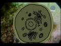
Ciclo Celular

Cuadro Sinóptico, tomado del blog de Hernán Darío Ramírez, url: http://ramirezrhernan.blogspot.com/
Nota: Corregir las impresiones ortográficas en Fases...çç
JC

MICROSCOPIO OPTICO

felicitaciones! Erika
Foto tomada del Blog de Erika Quiceno, http://erikaquiceno.blogspot.com/2009/09/tincion-de-gram-resumen.html
TINCIÓN DE GRAMM

Foto tomada del Blog de Erika Quiceno: http://erikaquiceno.blogspot.com/2009/09/tincion-de-gram-resumen.html

viernes, 2 de octubre de 2009
endotelinas sent you a video: "los 5 Reinos de los seres vivos"
| | help center | e-mail options | report spam |
| endotelinas has shared a video with you on YouTube: Clasificación de los seres vivos JC breve descripción de los 5 reinos en los que estan clasificados los seres vivos que forman parte de nuestra biodiversidad | |
| © 2009 YouTube, LLC 901 Cherry Ave, San Bruno, CA 94066 | |
endotelinas sent you a video: "El Reino de los Hongos"
| | help center | e-mail options | report spam |
| endotelinas has shared a video with you on YouTube: A manera de síntesis JC El Reino Fungi o Reino de los Hongos comprende varios tipos de "extraños seres" que no son plantas ni animales. No hacen la fotosíntesis, sino que necesitan alimentarse de materia orgánica que descomponen. Se reproducen por esporas. Hay especies perjudiciales y otras muy beneficiosas para el ser humano. | |
| © 2009 YouTube, LLC 901 Cherry Ave, San Bruno, CA 94066 | |

Micología
 Universidad Católica de Oriente - Facultad de Educación
Curso: Microbiología - Sesión * Docente: Esp. Juan Carlos González Sánchez
Octubre 3 de 2009 * Tópico: Micología
1. Generalidades.
2. Clasificación - Reproducción.
3. Micosis a nivel clínico
4. Aplicaciones
Actividades:
Revisar cada uno de los enlaces, en su portafolio o blog, presente un resumen – en el formato que prefiera – donde consigne una síntesis de la información revisada
1. Micología: Generalidades. http://www.slideshare.net/franciskoleon/generalidades-hongos
http://www.slideshare.net/furia/micologa-generalidades
2. Micología: Clasificación – Reproducción
http://200.93.199.34/areas_academicas/biology/multimedia/presentaciones/quintoquibio/hongos.ppt
www.uprm.edu/biology/cursos/biologiageneral/OClab5.ppt
COMPLEMENTARIO: http://mail.fq.edu.uy/~microbio/MGral/T2007/hongos1.pdf
3. Micosis a nivel clínico
http://hsjd08.files.wordpress.com/2008/03/04-generalidades-de-hongos.ppt
4. Micología – Aplicaciones
http://html.rincondelvago.com/algas-y-hongos.html Parte final del documento.
Teacher JC
Facultad de Educación Universidad Católica de Oriente
viernes, 18 de septiembre de 2009
Laboratorio - Observando bacterias al microscopio
Recuerden mañana traer bata de laboratorio, papel blanco, buena disposición y leer el protocolo de laboratorio que se encuentra en el vínculo:
http://www.revistaciencias.com/publicaciones/EElpZEVkykPMncqxmd.php
Que hemos revisado previamente....
Será una transferencia epistemológica maravillosa
JC
http://www.revistaciencias.com/publicaciones/EElpZEVkykPMncqxmd.php
Que hemos revisado previamente....
Será una transferencia epistemológica maravillosa
JC
domingo, 13 de septiembre de 2009


Suscribirse a:
Comentarios (Atom)
